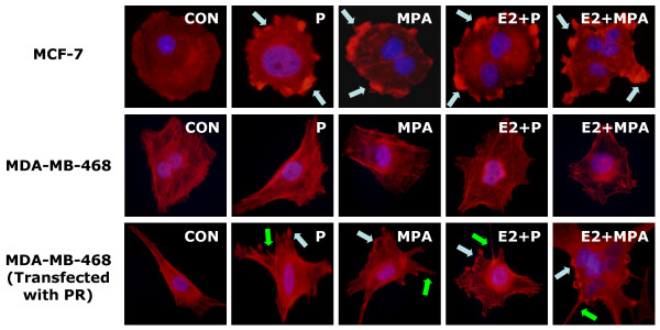
Figure 3.
MCF-7 cells and MDA-MB-468 cells with or without PR transfection were treated with P and MPA (both 100 nM), in the presence or absence of E2 (10 nM). Yellow arrows indicate longitudinal actin fibers, green arrows show pseudopodia, light blue arrows indicate ruffles. Nuclei are counterstained in blue.

