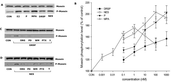
Figure 5.
Comparative effects of different progestin on moesin activation. (A) T47-D breast cancer cells were treated with E2 (10 nM) or progestins (P, MPA, DRSP all 100 nM; NES, 1 nM) for 15 min and total cell amount of Moesin or P-Moesin are shown. (B) Shows the concentration/effect curve of each progestin on moesin phosphorylation over a range of concentrations. * = P < 0.05 vs. DRSP at the same concentration. (C-D) Cells were exposed to DRSP (100 nM) or NES (1 nM) for 15 min, in the presence or absence of the pure PR antagonist ORG 31710 (ORG – 1 μM), of the MEK inhibitor PD98059 (PD – 5 μM) or of the PI3K inhibitor wortmannin (WM – 30 nM), of the G protein inhibitor, PTX (100 ng/mL) or of the ROCK-2 inhibitor Y-27632 (10 μM). Total cell amounts of Moesin or P-Moesin are shown.

