Fig. 3.
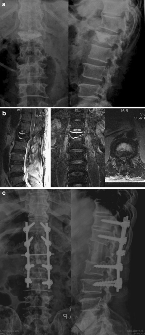
A 62-year-old man was diagnosed as infectious spondylitis clinically in conjunction with high values of erythrocyte sedimentation rate and C-reactive protein tests after vertebroplasty. a Radiography showed radiolucent lines between the bone and cement interface and L1 superior endplate erosion. b Sagittal and axial MRI demonstrated fluid (abscesses) surrounding the bone cement. c Anterior extensive debridement and reconstruction using a strut autograft, followed by posterior instrumentation was performed to control infection and restore spinal alignment and stability

