
Figure 3. Kinetics of cellular infiltration in lactate-supplemented Matrigel®.
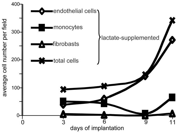
Differential cell counts were done on the basis of cellular morphology as identified by hematoxylin & eosin stain. From each implant, two sections were examined. Four fields per slide were examined at 40x magnification avoiding fields in which the periphery of the implant could be seen. Control slides, from implants either non-treated or treated with high molecular weight lactide polymer, showed too few cells to enable differential scoring. Clearly, lactate attracted primarily monocytes and endothelial cells.