
Figure 5. Histological characterization of Matrigel® implants in normoxic and hypoxic mice.
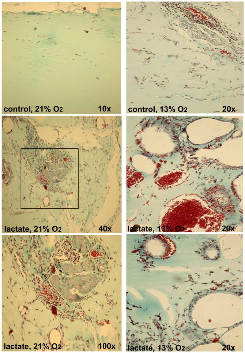
Thirty-six mice were each implanted with lactate-supplemented implant and one non-treated control. Twelve animals were held in each of three atmospheres: 13% (right column), 21% (left column), and 50% (not shown) oxygen for 11 days. A Pro-ox compact oxygen controller model 110 was used to maintain pO2 (Reming Bioinstruments, Redfield, NY). FiO2, humidity (60–80%), CO2 (<2 mm Hg) and temperature (23–25°C) were maintained within normal limits. Hypoxic (13%O2 ambience) were housed in a plexiglas chamber with a single gas inlet and outlet. Air was delivered at 1.0 L/min and nitrogen at 4.5 L/min from tanks connected to the chamber by thick-walled polyvinyl tubing. This reliably delivered 13.3 ± 1.3% oxygen to the chamber. Carbon dioxide levels were maintained at 0 mm Hg within the chamber with a CO2 absorber (Baralyme, Chemetron Medical Division, St. Louis, MO). A Datex Capnomac II [Datex Medical Instruments, Inc, Tewksbury, MA] was used to sample exhaust gas from the oxygen chambers to confirm the exposure pO2 and to ensure that CO2 did not accumulate. Compared to that in normoxic mice (left column), implants in hypoxic mice produced rare and large, thin-walled vessels. These photographs were selected to show as many vessels as possible and are not representative of their abundance. Compared to that in normoxic mice, small amounts of collagen were evident in implants in hypoxic mice. The sections were stained with Masson’s trichrome. For interpretation of the references to color in this figure legend, the reader is referred to the web version of this article at www.liebertonline.com/ars.