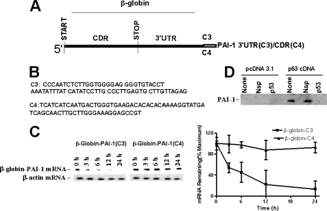
FIGURE 6.
Determining the destabilizing function of p53-binding PAI-1 3′-UTR mRNA sequence. A, structure of β-globin/PAI-1 chimeric mRNA. The p53 protein-binding 70-nt 3′-UTR sequence corresponding to nt 1958–2027 (C3) and a nonbinding control sequence of similar size corresponding to the coding region from nt 468 to 538 (C4) of PAI-1 cDNA were inserted into the 3′-UTR of β-globin cDNA. The chimeric β-globin/PAI-1 cDNAs were subcloned to pcDNA 3.1. B, nucleotide sequence of the p53 binding region nt 1958–2027 (C3) or nonbinding control sequence 468–538 (C4). C, decay of β-globin/PAI-1 chimeric mRNA. Beas2B cells were transfected with the chimeric β-globin/PAI-1 3′-UTR gene containing the 70-nt (nt 1958–2027) p53-binding sequence (β-globin-PAI-1(C3)) or nonbinding control sequence of PAI-1 CDR (nt 468–538) (β-globin-PAI-1(C4)) in pcDNA 3.1. Total RNA was isolated at different time intervals after treatment with DRB as described above (Fig. 3B) and analyzed for the level of chimeric mRNA by Northern blotting. Densitometric scanning of individual bands from four experiments is shown as a line graph. D, effect of p53 binding PAI-1 mRNA 3′-UTR sequence on PAI-1 expression. H1299 cells expressing vector cDNA (pcDNA 3.1) or p53 cDNA were untreated (None) or transfected with chimericβ-globin/PAI-1 cDNA containing a non-p53-binding control (C4, Nsp) or p53-binding (C3, p53) PAI-1 3′-UTR sequence in pcDNA 3.1, as described in Fig. 6C. PAI-1 expression in the CM was determined by Western blotting using an anti-PAI-1 antibody. The data shown are representative of the findings of four independent analyses.

