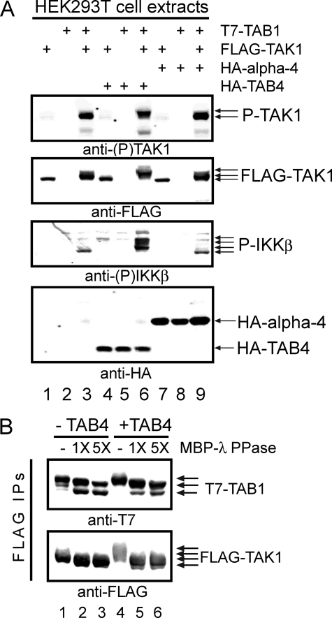
FIGURE 1.
TAK1 phosphorylation and activation by TAB4. A, combinations of FLAG-TAK1, T7-TAB1, HA-TAB4, and HA-α-4 were expressed in HEK293T cells and analyzed by immunoblotting with (top to bottom) 1) anti-(Thr(P)184/Thr(P)187) TAK1, 2) anti-FLAG, 3) anti-(Ser(P)177/Ser(P)181) IKKβ, and 4) anti-HA (bottom panel). Lanes 1–3 show extracts from cells co-expressing 1) FLAG-TAK1, 2) T7-TAB1, or 3) FLAG-TAK1 and T7-TAB1. Lanes 4–6 show extracts from cells co-expressing HA-TAB4 with FLAG-TAK1 and/or T7-TAB1. Lanes 7–9 show extracts from cells co-expressing HA-α-4 with FLAG-TAK1 and/or T7-TAB1. B, phosphatase treatment of FLAG-TAK1 and T7-TAB1. Proteins were co-expressed without (lanes 1–3) and with HA-TAB4 (lanes 4–6) in HEK293T cells, immunoprecipitated using anti-FLAG M2 beads, and immunoblotted with anti-T7 (upper panel) and anti-FLAG (lower panel). Immunoprecipitates (IPs) were treated with vehicle, 1×, or 5× amounts of recombinant MBP-λ-phosphatase (PPase) as indicated. P-, phospho-.

