
Fig. 6.
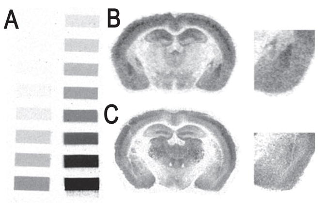
Representative bright-field photomicrographs showing GABAA receptor binding signals at the level of the amygdala in the mouse brain. Photomicrographs correspond roughly to coronal sections 1.4 mm posterior to Bregma as referenced by Paxinos & Franklin (2001). (A) Tritium standards. (B) [3H]-Flunitrazepam binding signal. (C) [3H]-Muscimol binding signal.