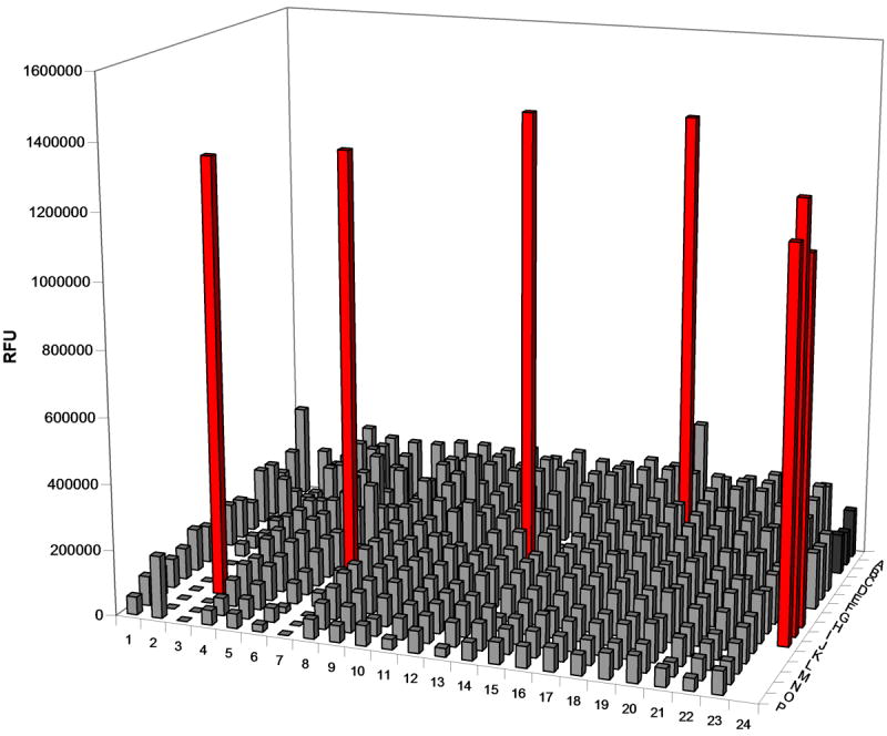
Figure 6.

High-throughput screen simulation. A 12.5 nM solution of the fluorescently-labeled oligonucleotide substrate was incubated in wells of a 384-well plate filled with either MazE:MazF(His)6 (1.5 μM:3.0 μM) or MazF(His)6 (3 μM). Control wells were prepared in column 24 in which oligonucleotide was incubated in the absence of compound with elution buffer (dark grey), MazE:MazF(His)6 (1.5 μM:3.0 μM; light grey), and MazF(His)6 (red). After a 2.5 hr incubation, wells containing MazF(His)6 (M3, J7, G13, and C18) are easily distinguished from those that contain MazE/MazF(His)6. These results demonstrate that the fluorescent oligonucleotide substrate could be used to detect MazEF complex disruptors in a high-throughput screen.
