
FIG. 1.
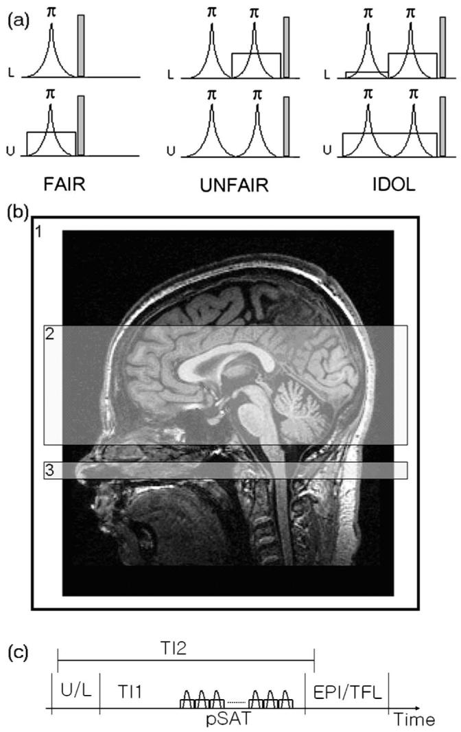
Three types of pulsed arterial spin labeling methods (a) with imaging position (b). (a) Labeled (L) and unlabeled (U) schemes of FAIR, UNFAIR, and the proposed method (IDOL). A radio frequency pulse was shown in a curve, representing the inversion pulses with flip angle of 180 deg. A spoiler gradient was applied after rf pulses to minimize a residual magnetization. A slice-selective gradient was represented by a rectangular shape. The amplitude of the first small gradient was decided by the size of the used head coil. (b) Locations of labeling by using an entire head coil (area 1) used in the first slice-selective gradient of the labeling (L) in the IDOL, imaging (area 2) used in other slice-selective gradient in all three methods, and the periodic saturation pulse (area 3). (c) The schematic drawing of the pulse sequence. pSAT is the periodic saturation pulses applied in area 3 in (b).