
Figure 8.
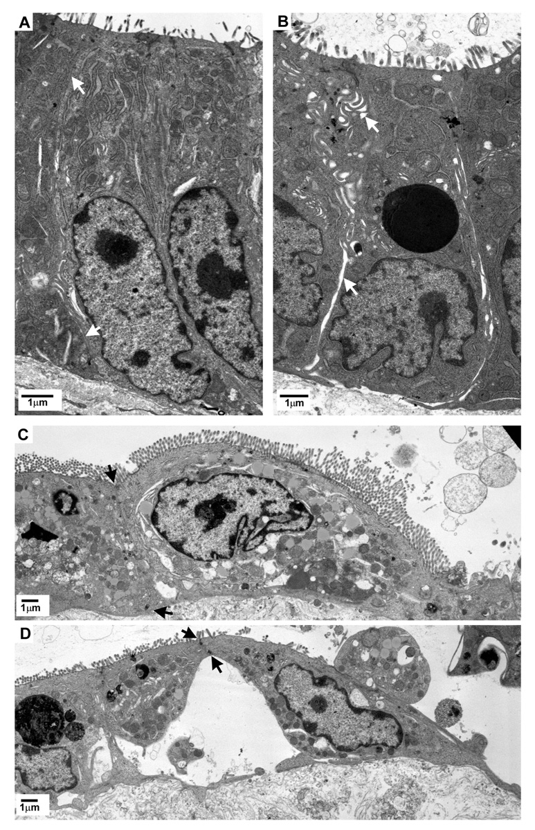
Transmission electron micrographs of crypt (A&B) and migrating villous enterocytes (C&D) from porcine ileal mucosa at peak repair after deoxycholate injury (time = 210-min). Mucosae shown in B & D were treated with indomethacin (INDO; 5×10⁻⁶M) immediately after injury and prior to onset of repair. White arrows indicate intercellular space. Black arrows indicate sites of lateral membrane attachment between adjacent, migrating enterocytes.