
Figure 7.
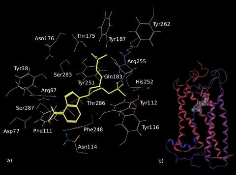
Model of the complex formed by MRS2179 and GPR17 after 3 ns of MD simulation. MRS2179 is displayed in yellow within the binding pocket in the detailed picture (a) and as spheres in the schematic representation of the entire ligand-receptor complex (b). In the representation of the whole receptor-ligand complex, helices and spheres are coloured as indicated in Figure 6.