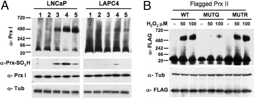
Fig. 4.
High-molecular-mass complexes in LAPC4 and LNCaP cells. (A) Native gels showing Prx I high-molecular-mass complexes induced by exposure to H2O2 for 20 min in LNCaP and LAPC4 cells. Concentration of H2O2 (lanes): 1, none; 2, 25 μM; 3, 50 μM; 4, 100 μM; 5, 100 μM + fluid change and recovery for 1 h. (Lower) Denaturing gels showing the levels of oxidized Prx (α-Prx-SO3H), Prx I (α-Prx I), and α-tubulin (α-Tub) in the LNCaP and LAPC4 lysates. (B) Flagged Prx II high-molecular-mass complexes in LNCaP cells treated with the indicated concentrations of H2O2; WT, and transfected with MUTQ (K196Q) or MUTR (K196R) are shown (see Results).

