
Figure 1.
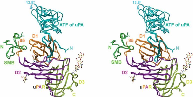
Recognition of both the uPA N-terminal fragment (ATF) and the vitronectin SMB domain by uPAR. Stereoview of superimposed crystal structures of the uPAR–ATF–SMB complex and the uPAR–ATF–SMB-antibody complex are shown (the antibody is omitted for clarity). The carbohydrate moieties of uPAR are shown in sticks. The three domains of uPAR are colored differently.