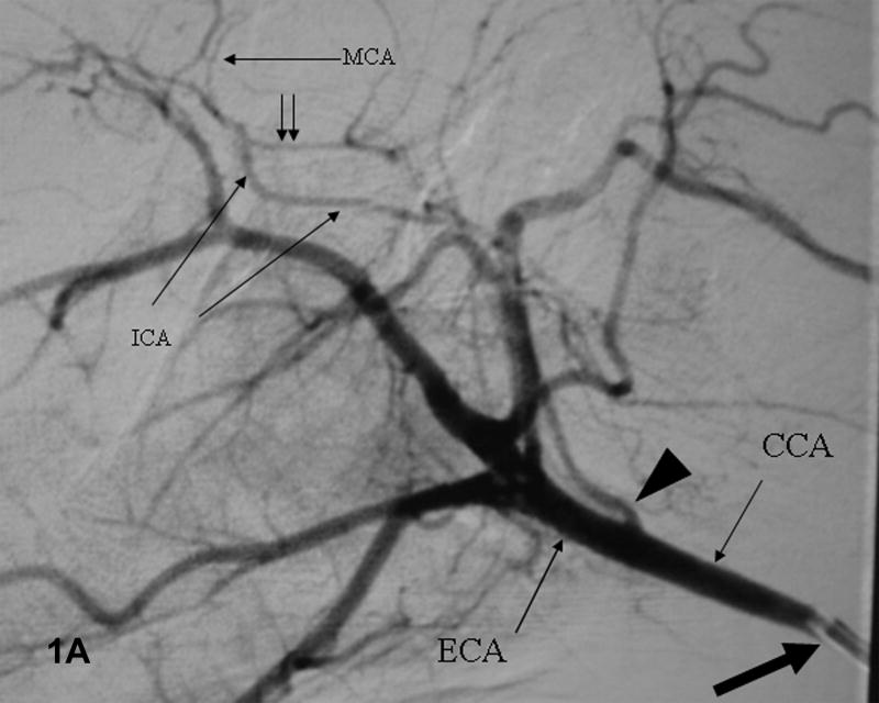
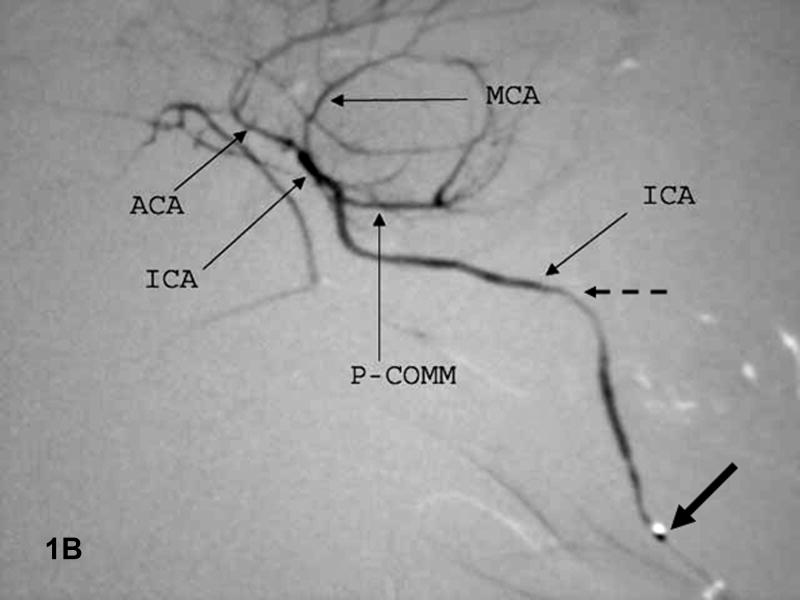
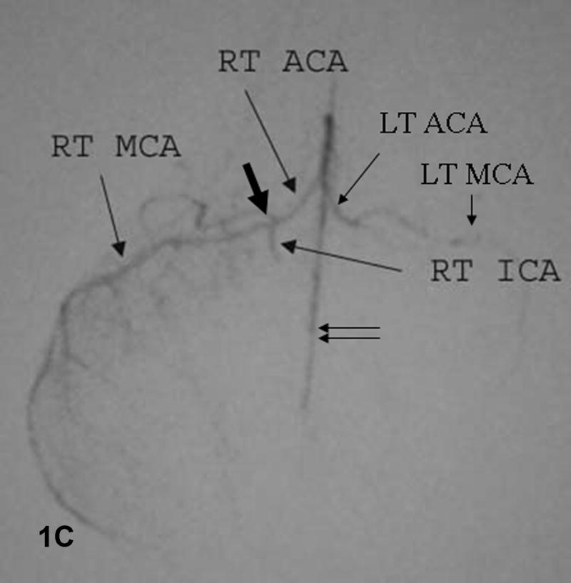
Figure 1. Baseline angiograms.
A. Lateral view. Catheter (thick arrow) is in the CCA. Arrowhead indicates the ICA take-off and double arrows the P-comm. B. Lateral view. The microcatheter tip (thick arrow) is in the ICA which narrows significantly at the skull base (dashed arrow). C. Submental vertex view. The microcatheter is in the ICA above the P-comm. ICA bifurcation into the MCA and ACA noted by the thick arrow, distal ACA by double arrows. The left ACA and MCA fill via the A-comm.


