
Fig. 1.
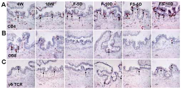
Immunohistochemistry for CD4 (A), CD8 (B), and γδ TCR (C) in the conjunctiva of NOD.B10.H2b mouse mice. Representative staining in 4-week-old untreated mice (4W), 16-week-old untreated mice (16W), and 16-week-old mice subjected to an air draft without (F) or with (FS) systemic scopolamine administration for 10 days (D). The CD4, CD8, and γδ TCR positive cells are indicted by arrows. Original magnification X400, bar = 25μm.