
Fig. 3.
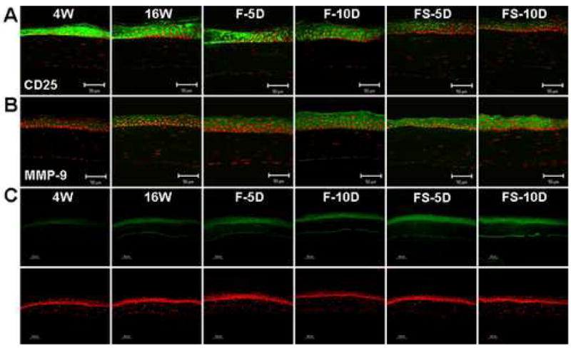
CD25 and MMP-9 expression and gelatinolytic activity in the corneal epithelium of NOD.B10.H2b mice. (A and B) Immunofluorescent staining and laser scanning confocal microscopy in corneal tissue sections stained with antibodies (green) to CD25 (A) and MMP-9 (B). (C) In situ zymogram showing gelatinolytic activity (green) in the corneal epithelium. Nuclei were counterstained with propidium iodide (red). Representative staining in 4-week-old untreated mice (4W), 16-week-old untreated mice (16W), and 16-week-old mice subjected to an air draft without (F) or with (FS) systemic scopolamine administration for 5 and 10 days (D). Original magnification X400, bar = 50 μm.