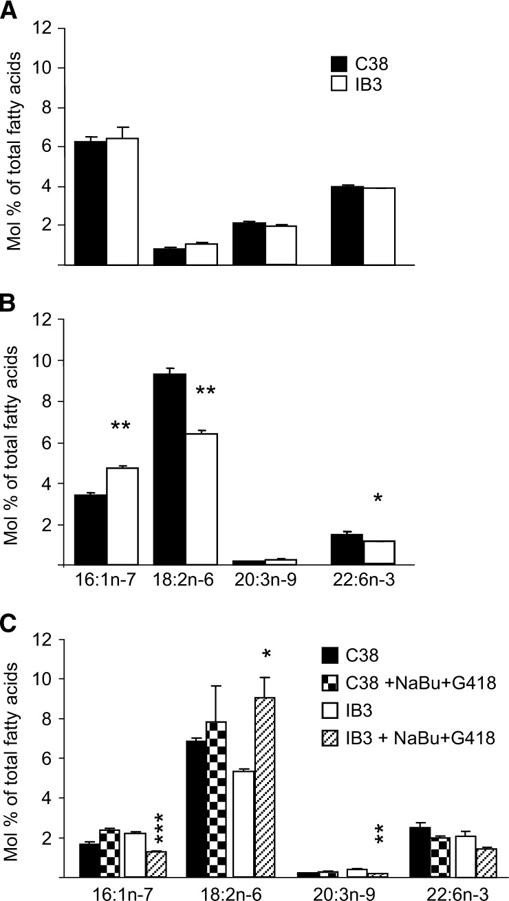
Fig. 3.
Fatty acid profile in C38/IB3-1 cells. C38 [wild-type (WT)] and IB3-1 (CF ΔF508/W1282×) cells were cultured in (A) 10% FBS, (B) 10% horse serum lot A, or (C) 10% horse serum lot B and incubated with and without sodium butyrate (2.5 mM) and G418 (150 μg/ml) for 48 h before harvest. Fatty acids were extracted, methylated, and analyzed by GC-MS. The internal standard was 17:0. Data are expressed as the mean ± SEM and are representative of a minimum of three different experiments, with each condition tested in triplicate. * P < 0.05, ** P < 0.01, *** P < 0.001.

