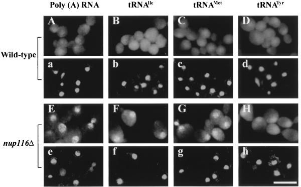
Figure 1.
Nuclear accumulation of tRNAs in nup116Δ cells. Parent W303 (A–D) and nup116Δ strain SWY27 (E–H) were grown at 23°C, and log phase cells were shifted to 37°C for 2 h. FISH was used to detect poly(A) RNA (A and E), tRNAIle (B and F), tRNAMet (C and G), and tRNATyr (D and H). a–h are 4′,6-diamidino-2-phenylindole staining of cells shown in A–H, respectively. (Bar = 10 μm.)

