Figure 4.
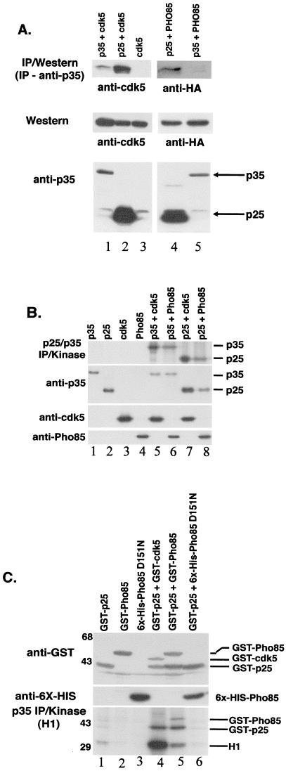
Activators of Cdk5 bind and activate Pho85 in mammalian and insect cells. (A) Coimmunoprecipitation of p35/p25 with Pho85. p35 antibodies were used to generate immunoprecipitates from lysates of COS-7 cells that had been transiently transfected with various combinations of CMV-p35, CMV-p25, CMV-cdk5, and CMV-HA-PHO85 as indicated at the top. Immunoblots were reacted by blotting with either anti-cdk5 (DC17) antibodies (Left) or anti-HA (12CA5) antibodies to detect HA-Pho85 (Right). Lanes: 1, p35+cdk5; 2, p25+cdk5; 3, cdk5 alone; 4, p25+PHO85; 5, p35+PHO85. Levels of Cdk5, HA-Pho85, and p25/35 expression in the extracts used in the immunoprecipitation reactions are shown (Middle and Bottom). (B) Phosphorylation of p25 and p35 by Pho85. Immunoprecipitation-kinase reactions were performed with anti-p35 antibodies by using lysates from COS-7 cells transfected with the following vector combinations: lane 1, CMV-p35; lane 2, CMV-p25; lane 3, CMV-cdk5; lane 4, CMV-HA-Pho85; lane 5, p35+cdk5; lane 6, p35+Pho85; lane 7, p25+cdk5; lane 8, p25+Pho85. The positions of migration of phosphorylated p35 and p25 are shown to the right. Levels of protein expression were confirmed by Western blotting with anti-p35, anti-cdk5, or anti-HA (Pho85) antibodies as indicated to the left. (C) Activation of Pho85 by p25 in insect cells. Lysates from Hi5 insect cells infected with baculoviral vectors expressing various combinations of GST-p25, GST-Pho85, 6XHis-Pho85D151N, and GST-Cdk5 were incubated with glutathione (GSH)-Sepharose beads to pull down the GST-tagged proteins. Kinase reactions were performed on the GSH precipitates with Histone H1 as substrate. The following proteins were present in the lysates: lane 1, GST-p25; lane 2, GST-Pho85; lane 3, 6XHis-Pho85D151N; lane 4, GST-p25+GST-cdk5; lane 5, GST-p25+GST-Pho85; lane 6, GST-p25+6XHis-Pho85D151N. The positions of migration of phosphorylated GST-Pho85, GST-p25, and Histone H1 (H1) are indicated to the right. Levels of protein expression in the lysates are shown by anti-6X-HIS Western blotting (Middle, to detect the Pho85D151N protein) and anti-GST blotting (Top).

