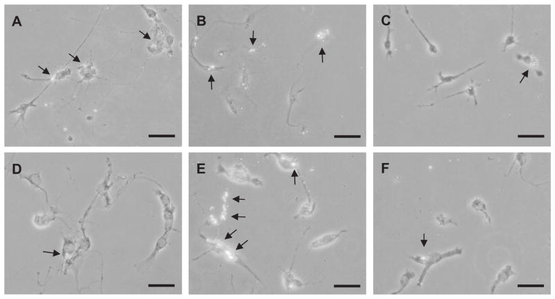
Fig. 5.
Detection of ingested Aβ42 in primary TLR4 wild-type and TlrLps-d/TlrLps-d microglia by fluorescent immunocytochemistry. Primary TLR4 wild-type (A–C) and TlrLps-d/TlrLps-d (D–F) microglia were treated with LPS (100 ng/ml) (A and D), CpG-ODN (0.51 μM) (B and E) or PBS (C and F) and exposed to oligomerized Aβ (0.25 μM) for 24 h. Ingested Aβ42 in the cells shows fluorescence by immunocytochemistry using 6E10 antibody and anti-mouse IgG antibody coupled with Alexa Fluor 488. Arrows indicate ingested Aβ. Scale bars 40 μm.

