
Fig. 6.
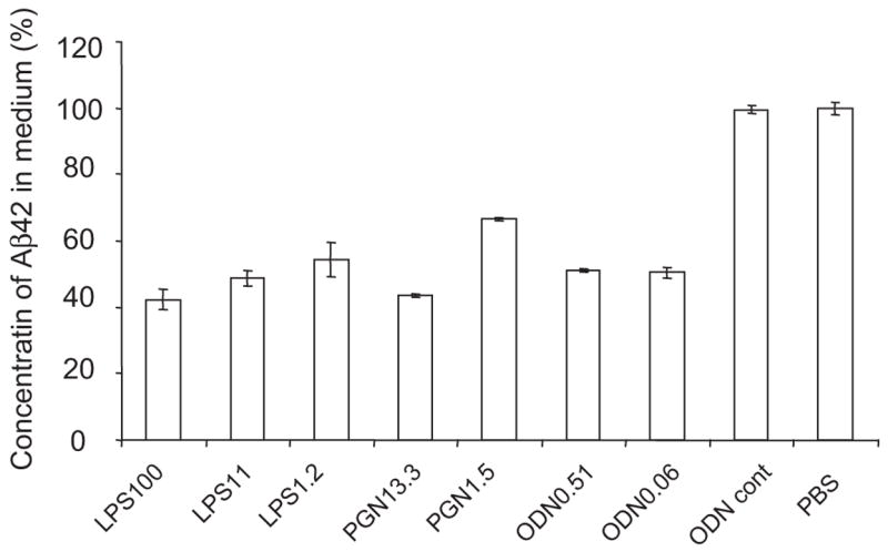
The clearance of Aβ42 from culture media by activation of TLRs on microglia (BV-2 cells). BV-2 cells were treated with LPS at the concentrations of 1.2, 11 and 100 ng/ml, CpG oligodeoxynucleotides (CpG-ODN) at 0.06 and 0.51 μM or peptidoglycan (PGN) at 1.5 and 13.3 μg/ml. BV-2 cells treated with the TLR ligands were incubated with oligomerized Aβ42 for 24 h. As controls, BV-2 cells treated with Control ODN or PBS were used. The concentrations of residual Aβ42 in the culture media were determined by Aβ42-specific sandwich ELISA. The ratios of the Aβ42 concentrations in TLR ligand-treaded media to that in PBS-treated medium are shown. After stimulation with every TLR ligand tested, residual Aβ42 in the medium was reduced by ~50% at the TLR ligand concentrations tested, compared with that in the media from BV-2 cells treated with PBS or control ODN (P < 0.01 for every TLR ligand treatment).