
Fig. 8.
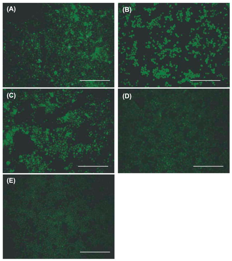
Detection of ingested Aβ42 in BV-2 cells by fluorescent immunocytochemistry. After treatment with LPS (100 ng/ml) (A), PGN (13.3 μg/ml) (B) or CpG-ODN (0.51 μM) (C), ingested Aβ42 in BV-2 cells shows fluorescence by immunocytochemistry using 6E10 antibody and anti-mouse IgG antibody coupled with Alexa Fluor 488. BV-2 cells treated with control ODN (D) or PBS (E) show little to no fluorescence. Scale bars 250 μm.