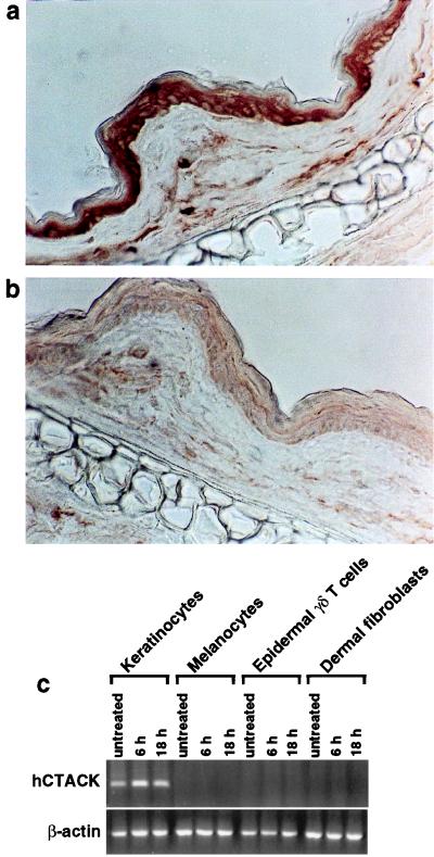
Figure 3.
Expression of CTACK. (a) Frozen sections of mouse skin (ears) were immunostained with a panel of anti-CTACK mAbs. A representative mAb (isotype IgG2b) is shown and revealed predominant staining in the epidermis and scattered positive cells in the dermis. (b) A control rat mAb (IgG2b) is shown for comparison. (c) RT-PCR analysis of CTACK expression in primary keratinocytes, primary melanocytes, 7-17 cells (epidermal γδ T cell line), and primary dermal fibroblasts either untreated or treated with TNF-α and IL-1β for the indicated times.

