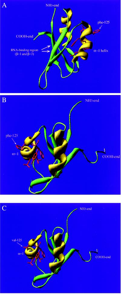
Figure 5.
Tertiary folding model of the immunodominant region of the U1-70K protein. β-sheets are presented in green, α-helices in yellow, and loops in light green. The NH3 and COOH ends of the fragment are depicted. (A) The immunodominant region with the RNA-binding β1- and β3-sheets as well as the α-helix-1 are depicted. Side chains of mutated amino acid residues in the 119–126 conformation-dependent epitope are presented in red (residues 119, 122, 123, 125, and 126). The arrow points to the amino acid residue at position 125. (B) The structure model viewed from the opposite direction. The 119–126 epitope is depicted with the Dm residue phenylalanine at position 125. (C) The epitope depicted with the human valine at amino acid residue 125.

