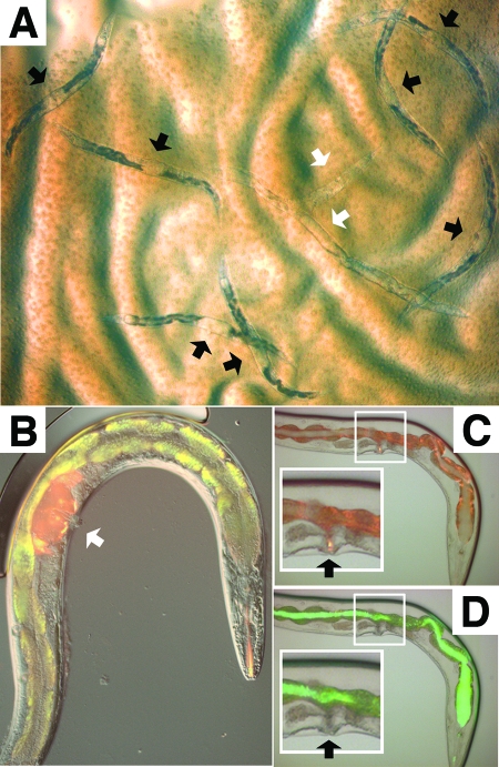
FIG. 1.
Leucobacter chromiireducens subsp. solipictus strain TAN 31504 accumulates within the uterus of C. elegans via the external vulval opening. (A) Bright-field micrograph of Glp adult worms feeding on a lawn of TAN 31504 after 5 days of exposure (×8 magnification). Black arrowheads point to the accumulated bacterial pathogen within the uterus of dead and dying worms. The white arrowheads indicate degraded worm carcasses. (B) Image of N3 filter (red) and I3 long-pass filter (green-red) fluorescence micrographs overlain on the corresponding DIC micrograph of a Glp adult worm exposed to TAN 31504 for 5 days followed by treatment with 10-kDa tetramethylrhodamine dextran (×200 magnification; images at ×400 magnification are shown in Fig. S1D to F in the supplemental material). The white arrowhead points to the accumulated red 10-kDa dye within the uterus of the infected worm. Autofluorescent granules, predominately of the worm intestine, appear as yellow in the merged image shown. The intensity of autofluorescence dramatically increases during C. elegans exposure to TAN 31504; the reason for this is unknown. (C) Image of N3 filter fluorescence micrograph overlain on the corresponding bright-field micrograph of a Glp adult worm exposed to S. enterica serovar Typhimurium SM022 for 5 days followed by treatment with 10-kDa tetramethylrhodamine dextran (×200 magnification; images at ×400 magnification are shown in Fig. S1A to C in the supplemental material). Fluorescence is predominately of the autofluorescent intestinal granules. The black arrowheads in panels C and D indicate the vulval openings. The insets, highlighted by white boxes, in panels C and D contain enlarged images of the vulva-uterus regions. The 10-kDa dye accumulated within the exterior vulval slit. (D) Corresponding I3 fluorescence micrograph overlain on the same bright-field micrograph depicted in panel C. The GFP-expressing pathogen accumulated within the worm intestinal lumen. The vulval slit was devoid of the GFP signal.

