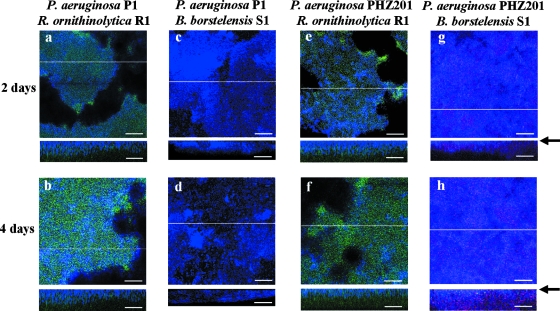
FIG. 3.
FISH analysis of biofilm on P. aeruginosa strain P1 biofilm in horizontal and vertical sections. Strain R. ornithinolytica R1 (panels a and b and panels e and f) or B. borstelensis S1 (panels c and d and panels g and h) was inoculated into a preestablished biofilm of strain P1 (a to d) or PHZ201 (e to h). P. aeruginosa P1 and PHZ201 appear blue with the Pae997-Cy3 probe, B. borstelensis S1 appears red with LGC-Cy5, and R. ornithinolytica R1 appears green with the ENT-FITC probe. Biofilms were cultivated for 2 or 4 days after inoculation into a preestablished biofilm of strain P1 or PHZ201. Vertical CLSM images show a cross-section through the x-z dimension at the positions marked by the lines on the horizontal panels. An arrow indicates an abiotic surface. Representative fields of vision are shown. Scale bars = 50 μm.

