FIG. 3.
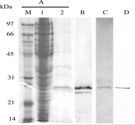
SDS-PAGE and Western immunoblotting of anti-CR1 scFv protein produced by E. coli. (A) Coomassie blue-stained SDS-PAGE gel. Lane M, low-molecular-mass protein markers; lane 1, crude cell lysate; lane 2, anti-CR1 scFv eluted from the NiCAM column. (B) Immunoblot of the isolated anti-CR1 scFv probed with the anti-HA antibody. (C) Coomassie blue-stained K8 scFv isolated by NiCAM chromatography. (D) Immunoblot of the isolated K8 scFv probed with the anti-HA antibody.

