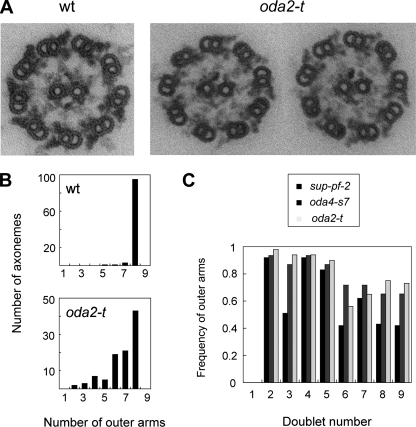
FIG. 5.
Electron microscopic analysis of the oda2-t axonemes. (A) Wild-type (wt) and oda2-t cross sections. Note that the outer-arm images for oda2-t appear thinner than for the wild type and are frequently missing or are weakened on some outer-doublets. (B) Number of the outer-doublet microtubules that bear outer arms in axonemal cross sections. (C) The average frequency of the presence of outer arms on particular outer doublets. The number of images used was 43 for oda2-t and 32 for oda4-s7. The data for sup-pf-2 are from Rupp et al. (38). In the wild-type axonemes, the outer doublet that lacks outer-arm dyneins is doublet no. 1, and other outer doublets are numbered clockwise in cross sections viewed from the base to tip direction (as in Fig. 5A). In the proximal region of the axoneme, the B tubule of doublets 1, 5, and 6 have beak-like structures (13), which serve as markers for numbering. These beaks are clearly seen in the two oda2-t images in panel A. In 25 wild-type images, the average numbers of outer arms were 0.0 for doublet 1 and 1.0 for all other doublets.

