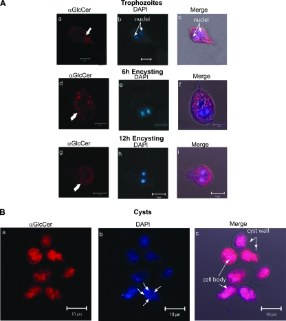
FIG. 5.
Stage-specific localization of GlcCer in Giardia. Nonencysting, encysting, and water-resistant cysts were labeled with monoclonal antibody against GlcCer, which was followed by reaction with TMR-conjugated rabbit anti-mouse antibody, as described in Materials and Methods. (A) Panels a and c show that in nonencysting trophozoites, GlcCer localizes to the plasma membranes. In 6- and 12-h encysting cells, the majority of the antibody labeling is localized in vesiclelike structures (panels d and f) and in the area surrounding the nuclei (panels g and i). (B) In water-resistant cysts (panels a to c), anti-GlcCer labeling is prominent in the cell body. Antigen-antibody reactions were considered to be specific because no labeling was observed with anti-GlcCer antibody pretreated with 30 μg/ml GlcCer (GlcCer-to-antibody ratio, 3:1). In contrast, glucose had no effect on antibody binding (not shown). “Merge” represents the colocalization of TMR, DAPI, and differential interference contrast microscopy images. αGlcCer, anti-GlcCer antibody. Bars = 5 μm (A) and 10 μm (B).

