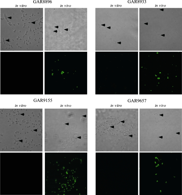
FIG. 5.
SdrG expression on S. epidermidis clinical isolates. Four groups of five mice each were infected with one of four MRSE clinical isolates (GAR8896, GAR8933, GAR9155, and GAR9657). At 3 hours postinfection, the blood from the mice was pooled and the bacteria isolated. The bacteria at the time of challenge (in vitro) and following isolation from the bloodstream (in vivo) were then stained and visualized as for Fig. 4. Arrowheads point to the bacteria in the bright-field images. Preimmune serum did not react with any of the bacteria grown in vitro or in vivo (data not shown). These pictures are representatives of three independent experiments.

