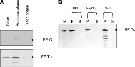
FIG. 5.
M. pneumoniae EF-Tu association with mycoplasma membrane. (A) Triton X-114 phase partitioning of intact M. pneumoniae cells. Equal amounts of sample were separated on Nu-PAGE gels under reducing conditions and transferred onto nitrocellulose membranes. Immunoblotting was performed with mouse anti-EF-Tu (1:3,000) or rabbit anti-EF-G (1:3,000) antiserum in 3% Blotto for 1 h at RT. (B) Alkali and high-salt treatment of M. pneumoniae membranes. M. pneumoniae S1 membranes (M) were treated with 3 M KCl, 0.1 M Na2CO3, and 1 M NaCl. Supernatant (S) and pellet (P) fractions were subjected to Nu-PAGE under reducing conditions, transferred onto nitrocellulose membranes, and immunoblotted with anti-EF-Tu antiserum as described in Materials and Methods.

