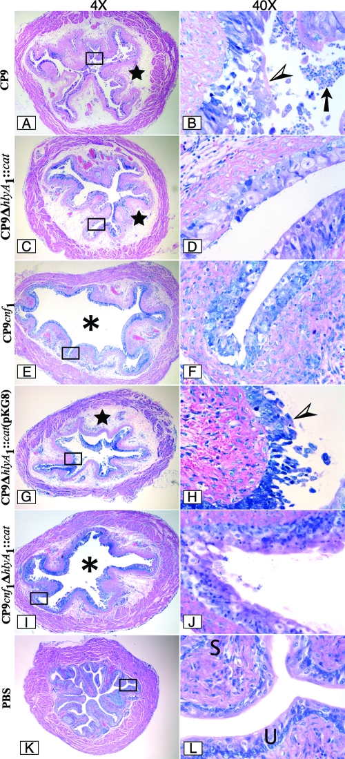
FIG. 6.
Infection of female C3H/HeOuJ mice with CP9 UPEC strains. Mice were inoculated with CP9 (A and B), CP9ΔhlyA1::cat (C and D), CP9cnf1 (E and F), CP9ΔhlyA1::cat(pKG8) (G and H), and CP9cnf1ΔhlyA1::cat (I and J) for 1 day, and then sections were cut, fixed with formalin, embedded in paraffin, stained with Giemsa stain, and analyzed by light microscopy. Images of a representative sample for each infection are shown at a magnification of ×4 on the left. The rectangles in the images indicate the sections that are shown at a magnification of ×40 on the right. The stars in panels A, C, and G indicate a high degree of edema. The asterisks in panels E and I indicate the luminal space. Note the presence of many inflammatory cells (arrow) in the luminal space in panel B. The arrowheads in panels B and H indicate the intense damage to the urothelium caused by CP9 and CP9ΔhlyA1::cat(pKG8), respectively. In contrast, the minimal damage caused by CP9ΔhlyA1::cat, CP9cnf1, and CP9cnf1ΔhlyA1::cat is shown in panels D, F, and J, respectively. Panels K and L show a murine bladder instilled with only PBS. U and S in panel L indicate the murine urothelium and submucosa, respectively.

