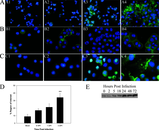
FIG. 4.
HMGB-1 is released from cells infected with Francisella both in vivo and in vitro. Immunofluorescence microscopy was performed to determine the localization of HMGB-1 in the lungs of infected mice and in BALF cells, as well as in infected J774 cells. HMGB-1 was visualized with Alexa 488 (green), while DAPI was used to observe the nuclei. Magnification for panels A1 to A4 and B1 to B4, ×800X; magnification for panels C1 to C4, ×1,000. (A) Images of similar areas in the lung (lung parenchyma and alveolar epithelium). Panel A1 is an image for the lung of a representative mock-infected mouse, while panels A2, A3, and A4 are images for mice infected with F. tularensis subsp. novicida at 6 hpi and 1 and 3 dpi, respectively. Panel A4 shows HMGB-1 expression in a lesion in the lung at 3 dpi. (B) HMGB-1 expression in cells isolated from the BALF. Panel B1 is an image for cells harvested from a mock-infected animal, while panels B2, B3, and B4 are images for cells harvested at 6 hpi and 1 and 3 dpi, respectively. (C) Representative micrographs of J774 cells infected with F. tularensis subsp. tularensis Schu S4 at an MOI of 50 at 0, 8, 24, and 72 hpi (panels C1 to C4, respectively). (D) Percentage of the region of interest positive for HMGB-1 in multiple tissue sections as determined by using IPLabs 3.7. Two asterisks, P < 0.01. (E) Representative Western blot for two independent experiments probed for the presence of HMGB-1 using equivalent amounts of whole-cell extracts of J774 cells infected with Schu S4 at an MOI of 50 at the following time points: 0 (mock infection), 2, 5, 18, 24, 48, and 72 hpi.

