FIG. 5.
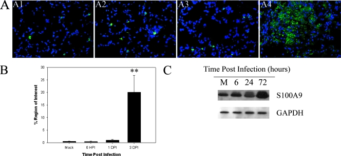
S100A9 expression is increased in the lungs of mice infected with F. tularensis subsp. novicida. (A) S100A9 expression assessed using immunofluorescence microscopy with lung sections from mice infected with F. tularensis subsp. novicida. Panel A1 shows S100A9 expression in mock-treated animals. Panels A2, A3, and A4 show S100A9 expression in F. tularensis subsp. novicida-infected lungs at 6 hpi and 1 and 3 dpi, respectively. All images are images of similar areas of the lung (lung parenchyma and alveolar epithelium). Magnification for all panels, ×400. (B) Percentage of the region of interest expressing S100A9 obtained from in situ immunofluorescence data for the lungs of mice infected with F. tularensis subsp. novicida using IPLabs 3.7 software. Two asterisks, P < 0.01. (C) Western blot demonstrating expression of S100A9 in lung homogenates obtained from mock-infected animals (lane M), as well as animals infected for 6, 24, and 72 hpi. The position of glyceraldehyde-3-phosphate dehydrogenase (GAPDH) is shown as a loading control for the assay. The blot is a representative Western blot for two independent experiments.

