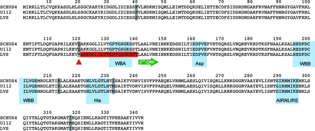
FIG. 1.
Analysis of pilT ORFs from F. tularensis SchuS4 (F. tularensis subsp. tularensis; FTT0088), U112 (F. tularensis subsp. novicida; FTN_1622), and LVS (F. tularensis subsp. holarctica; FTL_1770 and FTL_1771). The amino acid sequences of the ORFs are aligned, with nonconserved residues boxed in gray. The red triangle marks the location of the stop codon mutation in the LVS. The stop codon is followed by a stretch of 20 residues predicted not to be translated (boxed in red), which is followed by the predicted start site for the second ORF, indicated by the green arrow. The locations of the Walker box A (WBA), aspartate box (Asp), Walker box B (WBB), histidine box (His), and AIRNLIRE motifs are indicated. The motifs were identified by visual inspection of the PilT sequences and manual alignment with the motifs as previously defined in related family members (3, 54).

