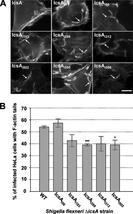
FIG. 5.
F-actin comet tail formation by intracellular S. flexneri ΔicsA strains expressing IcsA-i mutants. (A) IF microscopy of F-actin tail formation by intracellular S. flexneri ΔicsA strain expressing IcsA-i mutants. HeLa cells infected with S. flexneri were labeled with anti-LPS antibodies and Alexa 594-conjugated donkey antirabbit antibodies, and F-actin was labeled with FITC-phalloidin. Arrows indicate F-actin tail formation. Strains were assessed in three independent experiments. Scale bar = 10 μm. (B) Frequency of F-actin tail formation by S. flexneri ΔicsA strains expressing IcsA-i mutants. HeLa cells were infected with S. flexneri strains and examined by IF microscopy as detailed in Materials and Methods. The frequency of F-actin tail formation was determined by observing the percentage of infected HeLa cells (n = 100) that had at least 1 F-actin tail. Data represent means ± standard errors. *, P < 0.05; ***, P < 0.001 (determined by Student's unpaired two-tailed t test). Data are from three independent experiments.

