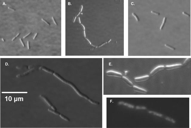
FIG. 5.
Differential interference contrast microscopy of Lud135/BC201. Cells were grown at 30°C in liquid LB medium to mid-log phase and visualized using a Nikon Microphot-FXA phase-contrast microscope. (A) Parent strain W3110A. (B) Lud135/pACYC184. (C) Lud135/p-yghB. (D) BC201. (E) Lud135/pACYC184 (close-up). (F) DAPI-stained Lud135/pACYC184 illustrating nucleoid segregation. Lud135 cells grown briefly at 42°C display a similar phenotype (not shown).

