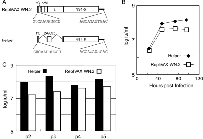
FIG. 6.
Analysis of two-component genome replication. (A) Schematic representation of RepliVAX WN.2 and helper genome structure showing the position and composition of the WT (RepliVAX WN.2) and mutant (helper) CS. Nucleotide substitutions at 5′ CS and 3′ CS are in lower case. (B) Release of infectious particles containing the genome of RepliVAX WN.2 or helper from Vero cells infected with a mixture of particles at an MOI of 0.5. The titers of particles recovered at each time point were determined by an infectious-unit assay performed on Vero cells (see Materials and Methods). The titer of the RepliVAX WN.2 particles was determined by focus formation assays using an E protein-specific serum that detects only cells infected with the E-encoding RepliVAX WN.2 genome. The titer of helper particles was determined by subtraction of the focus formation assay titer of RepliVAX WN.2 from the combined titer of RepliVAX WN.2 and helper particles determined by using an anti-WNV serum (see Materials and Methods). (C) Titers of infectious particles containing the genome of RepliVAX WN.2 and helper after serial passage in Vero cells. Supernatants collected 3 days postinfection were used to inoculate fresh Vero cells. This procedure was repeated five times, and the titers of both helper and RepliVAX WN.2 in each passage were determined as described for panel B.

