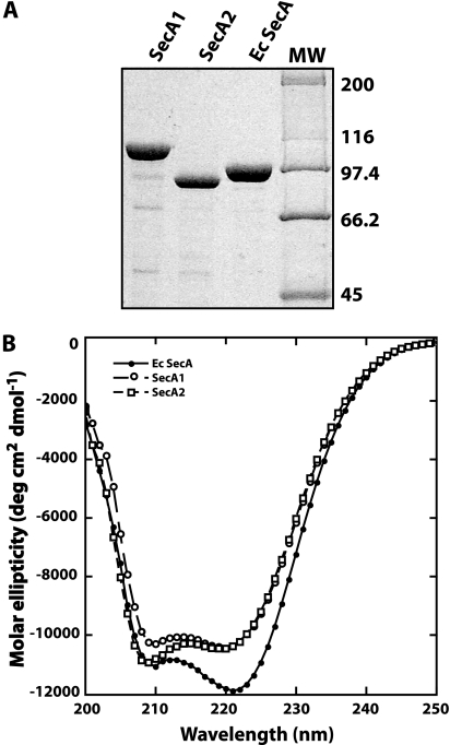
FIG. 1.
(A) SDS gel of the purified SecA proteins. SecA1, SecA2, and E. coli SecA were purified as described in Materials and Methods. The proteins were separated on a 10% SDS polyacrylamide gel. Molecular weight markers (MW, in thousands) are indicated on the right side of the gel. (B) CD spectra of each SecA protein. The spectra were obtained as described in Materials and Methods. The protein concentration was 0.1 mg/ml in 20 mM phosphate buffer (pH 7.6).

