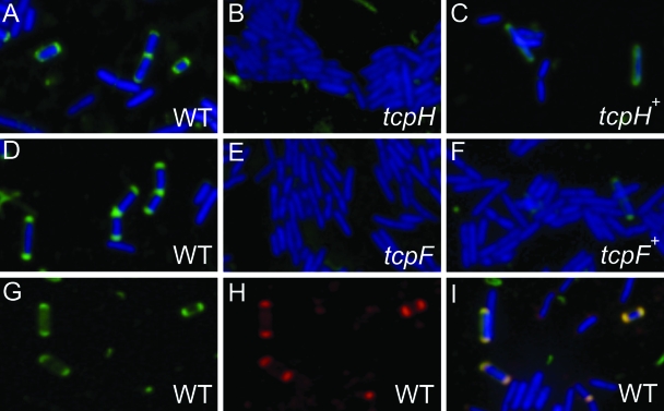
FIG. 10.
Localization of TcpH and TcpF by immunofluorescence microscopy. C. perfringens cells were labeled nonspecifically using the DAPI nuclear stain (blue), specifically using TcpH-specific (A to C) and TcpF-specific (D to F) primary antibodies, and corresponding Alexa Fluor 488(Green)-conjugated anti-guinea pig and anti-rabbit secondary antibodies, respectively. TcpH and TcpF were identified as green fluorescent polar foci. The C. perfringens strains analyzed were JIR325(pCW3) (A, D, and G to I), the tcpH mutant JIR4885 (B), the complemented tcpH mutant JIR4891 (C), the tcpF mutant JIR4940 (E), and the complemented tcpF mutant JIR4942 (F). For the colocalization of TcpF and TcpH by immunofluorescence microscopy, JIR325(pCW3) cells were probed concurrently with (G) TcpH-specific primary antibody and Alexa Fluor 488 (green)-conjugated anti-guinea pig secondary antibody and (H) TcpF-specific primary antibody and Alexa Fluor 568 (red)-conjugated anti-rabbit secondary antibody. The green fluorescent polar foci are TcpH specific, and the red fluorescent polar foci are TcpF specific. (I) Merged image of panels G and H and simultaneously DAPI stained cells.

