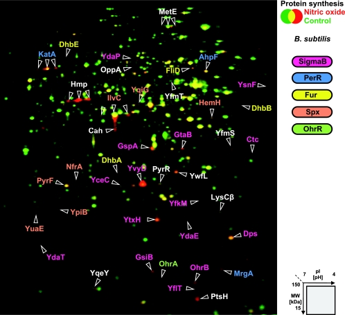
FIG. 2.
Synthesis of cytoplasmic proteins of B. subtilis after NO stress. The 2D gel images of newly synthesized proteins (labeled with l-[35S]methionine) from exponentially growing cells (shown in green) and cells exposed to 100 μM concentrations of the NO donor MAHMA NONOate for 10 min (shown in red) were overlaid. Identified proteins with an increased synthesis rate 1 to 30 min after stress are labeled and color coded for their membership to specific regulons as indicated.

