FIG. 4.
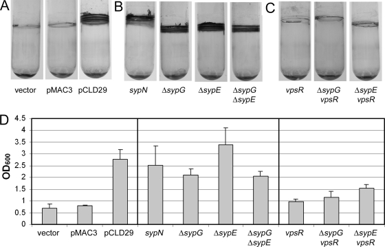
Crystal violet staining of glass-attached material following static growth. Glass-attached materials following growth of cells under static conditions were assayed by subculturing into 2 ml HMM with Tc and incubating at room temperature for 48 h and then staining with 1% crystal violet. (A) Crystal violet staining of glass-attached material from cultures of vector-, pMAC3-, and pCLD29-containing cells. (B) Crystal violet staining of glass-attached material from cultures pCLD29-containing syp mutants. (C) Crystal violet staining of glass-attached material from cultures of pCLD29-containing vpsR mutant and vpsR syp double mutant strains. (D) Quantification of the crystal violet-stained material from tubes shown in panels A to C (and additional tubes not shown), solubilized as described in Materials and Methods. Error bars represent standard deviations.

