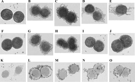
FIG. 4.
DAF and α5β1 integrin binding properties of DraE and DraD examined by light microscopy of E. coli strains stained with Giemsa stain. Adherence of E. coli strains to HeLa cells. (A to E) Microscopic examination of E. coli BL21(λDE3) (negative control of cellular adhesion) (A), E. coli BL21(λDE3)-pCC90 (positive control of cellular adhesion) (B), E. coli BL21(λDE3)-pCC90DraDmut (C), E. coli BL21(λDE3)-pCC90D54stop (D), and E. coli BL21(λDE3)-pCC90DraCmut (E). (F to J) Micrographs of E. coli BL21(λDE3) with the gspD gene knockout (GspD− strain) transformed with the same plasmids as shown in panels A, B, C, D, and E, respectively. (K to O) Micrographs of E. coli BL21(λDE3) GspD− mutant strain complemented with plasmid-encoded GspD and harboring the same plasmids as shown in panels A, B, C, D, and E, respectively. Forty cells associated with bacteria were examined by light microscopy. Magnification, ×10,000 (Olympus BX-60 microscope).

