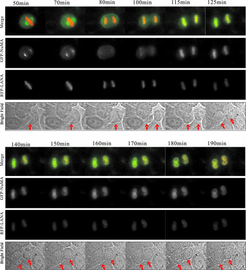
FIG. 5.
NuMA and LANA reunite in daughter nuclei after cytokinesis. U2OS cells were transfected with GFP-NuMA and RFP-LANA and synchronized at mitosis. Cells released from treatment continued to grow in a chamber in L15 medium supplemented with 10% fetal bovine serum. U2OS cells were filmed on a Leica inverted microscope equipped with a 63× 1.4-NA PlanApo objective lens. Images were recorded at 5-minute intervals and analyzed with Image J software. Selected frames from the two-color time-lapse recording are shown. Bright field images are shown under the merged images. Arrows in Phase panels indicate the cells focused for the localization of GFP-NuMA and RFP-LANA.

