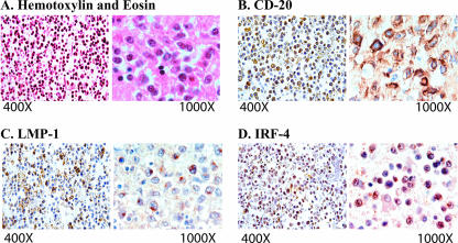
FIG. 5.
Histological and immunohistochemical characterization of IRF-4 in CNS lymphomas. (A) Hematoxylin and eosin staining of a CNS lymphoma demonstrated the perivascular concentric accumulation of neoplastic cells. (B) Immunohistochemistry for the pan-B-cell marker CD20 highlighted the neoplastic lymphocytes in the Virchow-Robin space and confirmed their B-cell origin. (C) The EBV LMP-1 was robustly expressed in the cytoplasm of neoplastic lymphocytes in both the perivascular space and the brain parenchyma. (D) The expression of IRF-4 was detected in numerous neoplastic lymphocytes.

