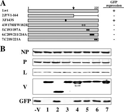
FIG. 4.
Repression of GFP expression from the minigenome system by mutant hPIV2 V proteins. (A) Schematic diagram of the V proteins. The closed circle indicates the position of the mutation residue of Phe143. The asterisks indicate the positions of the mutation residues of Trp-motif. The closed squares indicate the positions of the mutation residues of the Cys motif. The arrow above marks the editing site. (B) Effects of mutant V proteins on the GFP expression in the minigenome system. BSR T7/5 cells were transfected with plasmids pPIV2-GFP (1 μg), pTM1-NP (0.75 μg), pTM1-P (0.4 μg), and pTM1-L (0.75 μg) plus various pTM1-V (1 μg). After 48 h, the cells were assayed by Western blotting with anti-NP, P/V, L, and GFP antibodies. The numbers on the bottom of the figure correspond to each V protein described in panel A.

