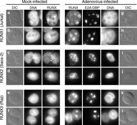
FIG. 3.
Human RUNX proteins accumulate at viral replication centers at late times during adenovirus infection. Jurkat cells, SaOS-2 cells, and Raji cells were either mock infected (a to f) or infected with the wild-type virus dl309 (g to l). After 24 h, the cells were extracted with Triton X-100 as described in Materials and Methods. Viral replication centers were visualized with the mouse monoclonal antibody B6-8 against the E2A-DNA-binding protein, and the RUNX protein was simultaneously visualized with rabbit polyclonal antibodies specific for RUNX1 (a, b, g, and h), RUNX2 (c, d, i, and j), or RUNX3 (e, f, k, and l). DNA was visualized by being stained with DAPI. The bar in each differential interference contrast (DIC) image represents 10 μm.

