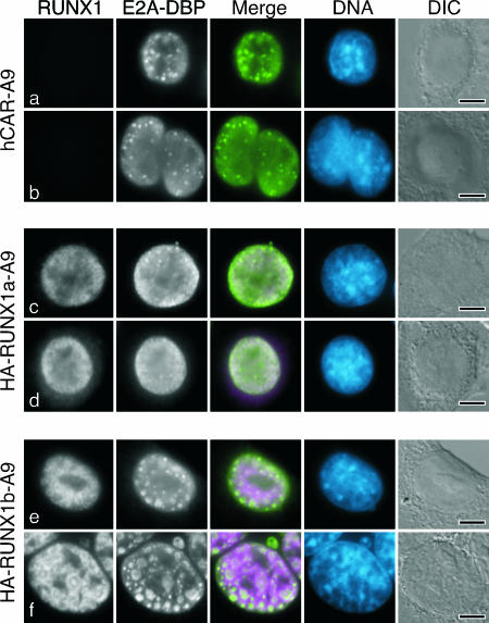
FIG. 9.
E1B-55K supports the formation of virus replication centers, and RUNX1b can compensate for the loss of E1B-55K in adenovirus-infected mouse cells. The hCAR-transduced mouse A9 cells analyzed in Fig. 5 were infected with the E1B-55K mutant virus dl1520 and processed for double-label immunofluorescence as intact cells after 24 h using the E2A-DBP-specific mouse monoclonal antibody B6-8 and the HA-specific rat antibody. In the merged image, E2A-DBP is shown in green and RUNX1 is shown in magenta. DNA was visualized by being stained with DAPI and is shown in blue. The bar represents 5 μm. DIC, differential interference contrast.

