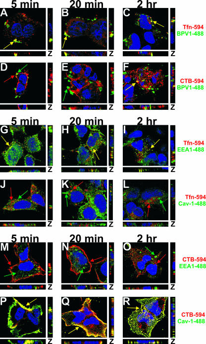
FIG. 1.
BPV1 PsVs colocalize with Tfn early during infection and later show overlap with CTB. (A to F) BPV1 PsVs were internalized into 293 cells simultaneously with 594-labeled Tfn (A to C, red) or CTB (D to F, red). Virions were labeled with anti-L1 antibody 5B6 (green), and TOPRO-3 was used to stain the nucleus (blue). At 5 and 20 min, the virus overlapped with Tfn (A and B, yellow) but not CTB (D and E). Two hours postinternalization, there was prominent overlap between BPV1 and CTB (F, yellow) and residual overlap with Tfn (C). (G to L) 293 cells were allowed to internalize Tfn (red) for 5 min, 20 min, and 2 h and stained for the early endosome marker EEA1 (G to I, green) and caveolin-1 (Cav-1) (J to L, green). Tfn colocalized with EEA1 at all time points (G to I, yellow). No overlap was seen between Tfn and caveolin-1 at any of the time points (J to L). (M to R) 293 cells internalized CTB (red) for 5 min, 20 min, and 2 h. EEA1 (M to O, green) and caveolin-1 (P to R, green) were labeled in the cells. Colocalization was observed between CTB and caveolin-1 at all time points (P to R, yellow) but not between CTB and EEA1 (M to O). Images shown are z stacks, representing the cell in the x, y, and z planes.

